Computer & Phantom Simulations of Lungs
- For human subject simulations, a 3-dimensional (3D) computational model for simulating the vibratory response of the human upper torso is developed by using the CT image sets from the Visible Human Male (VHM) of the National Library of Medicine. The dimension of the human torso model was rescaled to the approximate size of the human subjects (HS) experiments.
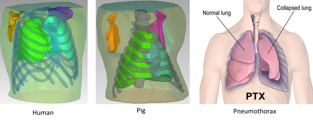
- For porcine model simulations, a pig of similar size to those studied in the acoustic experiments was scanned by an x-ray CT in 1 mm slice steps with pixel matrix size 512×512 (Brilliance 64, Philips Electronics). In both human and porcine studies, the CT image sets are then imported into Mimics V14 (Materialise, NV) which is an image processing software for 3D design and modeling, to generate the geometries of the 3D human and porcine models shown in the figures.
- The soft tissue, ribcage, sternum, scapula and lungs were detailed to create an accurate geometric model. Material property values used for the different tissue regions were based on previous studies of the authors, including [3], which details an experimental determination of the parameter values for a viscoporoelastic model of the lungs based on Biot theory. Viscoelastic values for other soft tissue regions and for bone can be found in [17].
- Harmonic vibratory excitation with displacement amplitude of 1 mm was applied on the sternum. The simulations were performed in a finite element (FE) environment COMSOL® 4.3 using the harmonic analysis acoustic-solid interaction module.
- To simulate the pathological condition, the original right lung region of the 3D model was separated into two parts (showing in the figure), the collapsed lung parenchyma region and air region. Two different levels of collapse were considered, which resulted in ∅=28% and ∅=58%. Those two values, respectively, correspond to PTX by volume percentages of 89% and 53%. The compression wave speed and attenuation used as the material properties, respectively, calculated by Biot theory for lungs at ∅ = 75%, ∅= 58%, and ∅= 28% based on [3].
Human Trials | Porcine Trials | Comparison to Computational & Phantom Simulations
Human Subjects


FRF vs. Frequency Plots

FRF of points #15(a), #24(b), and #41(c) on posterior chest (left/right sides correspond to left/right columns).

Comparison of normal and PTX states at point #41.


