Pig Percussion
Sound Transmission in Pig Lung by Percussion Heading link
Sound Transmission in Pig Lung by Percussion
Measuring sound transmission through the torso and lungs may be of value if altered transmission patterns correlate with pathology in ways that can be detected and used to provide a reliable and quantitative diagnosis of disease or injury. An accurate computer simulation of the sound transmission may aid in interpreting experimental measurements.
In the present study, vibratory excitation in the frequency range of 50-400 Hz is implemented on the chest surface of two normal porcine subjects. This technique introduces mechanical waves of broad frequency content that is more controllable than the low frequency content obtained by tapping in conventional auscultatory percussion. The underlying hypothesis of the technique is that the high-frequency mechanical waves generated by the vibratory excitation on the anterior chest propagate to the posterior chest through internal organs between the location of measurement and the point of excitation. The change in intrathoracic organs would partially alter the wave propagation and the resulting motion amplitude at the measurement location. By applying the vibratory excitation to the sternum, the posterior torso surface motion excitation was experimentally measured and used to validate the results obtained from a computer simulation of the vibratory response of the torso to the external excitation source. This computer simulation model is then used to predict changes in acoustic transmission caused by pneumothorax (PTX). Experimental measurements are also conducted on porcine subjects under PTX conditions. Similar trends in measured sound transmission and how it is affected by PTX are observed. The developed computational simulation models may be of use in assessing the performance of other acoustic approaches and the diagnosis of other injuries and diseases.
Experiment Setup Heading link
Experiment Setup
- Experiments were carried out on two freshly sacrificed female Landrace and Yorkshire cross pig subjects (PS) that weighed from 30 to 35 kg. Immediately upon sacrifice the lung was inflated by air with positive pressure of 5 cm H2O gage.
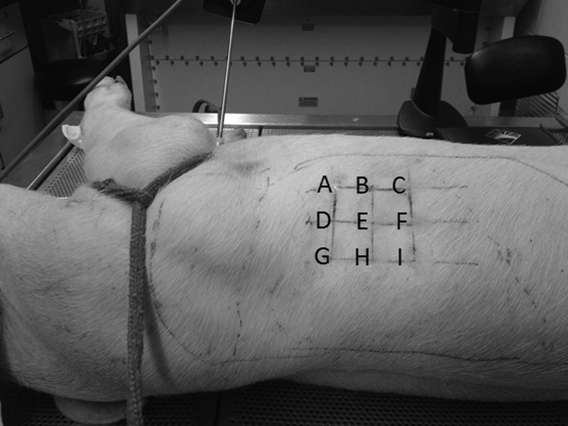
- A plexiglass disk of radius 15 mm was gently pressed against the chest surface on the top of the sternum and driven by an electromagnetic shaker (ET-132, Lab-Works Inc., Mesa Costa, CA) that was connected to a power amplifier (P 3500S, Yamaha, Buena Park, CA). The vibratory excitation has a spectral content from 50 – 400 Hz.
- The skin surface motions of 9 evenly spaced points in an array located in the area from the fifth rib to the ninth rib were measured by a laser Doppler vibrometer (LDV) (PDV-100, Polytec, Irvine, CA).
- After the “normal case” experiment was complete, a small incision was made in the seventh intercostal space in the right mid-axillary line to create the PTX state. A 5 mm thoracoscopic trocar (Endopath Dilating Tip, model 355, Ethicon, Cincinnati, OH) was inserted into the pleural space to confirm the PTX state.
- Then air was introduced through the incision using a syringe. The same measurements were taken under PTX state.
Simulations Heading link
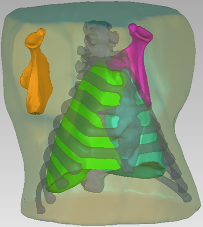
3D geometry of the pig upper torso
Simulations
- For porcine model simulations, a pig of similar size to those studied in the acoustic experiments was scanned by an x-ray CT (Brilliance 64, Philips Electronics) in 1 mm slice steps with pixel matrix size 512×512. The CT image sets are then imported into an image processing software Mimics V14 (Materialise, NV) to generate the 3D geometry of the pig upper torso.
- The soft tissue, ribcage, sternum, scapula and lungs were separated to create an accurate geometric model. Material property values used for the different tissue regions were based on previous studies.
- Harmonic vibratory excitation with displacement amplitude of 1 mm was applied on the sternum. The simulations were performed in a finite element (FE) software COMSOL® 4.3 using the harmonic analysis acoustic-solid interaction module.
- To simulate the pathological condition, the original right lung region of the 3D model was separated into two parts, the collapsed lung region and air region.
Results of Porcine Experimental and Simulation Studies Heading link
Results of Porcine Experimental and Simulation Studies
